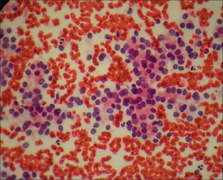
Figure 2.

Guided aspirate showing highly cellular smear with stippled nuclear chromatin and moderate pleomorphism. Cells are arranged predominantly in cohesive groups with many single cells/naked nuclei. Absence of colloid and macrophages noted (Pap, ×400)
