
Figure 4.
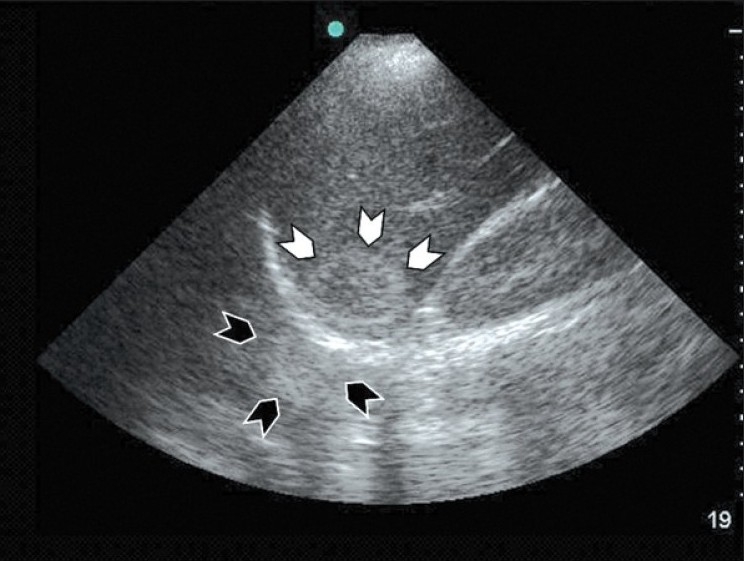
Coronal section of the liver using a curvilinear probe showing a haemangioma under the dome of the diaphragm (white arrow heads) and its mirror artifact above the diaphragm (black arrow heads). Notice that the mirror artifact is more blurred and distorted than the image of the original hemangioma