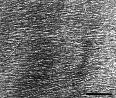
Figure 8.
Electron micrograph showing the appearance of microfibrils on the innermost layer of a longitudinal-radial cell wall of a cortical cell from a well-watered root. Image shows a cell with a net transverse orientation of microfibrils, approximately 5 mm from the apex. The longitudinal axis of the root is parallel to the side of the figure. Vibratome sections were extracted with carbonate and a metal-carbon replica was made as described in Methods. Bar = 400 nm.

