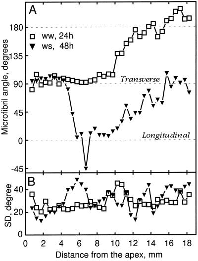
Figure 9.
Microfibril orientation as a function of distance from the apex of well-watered (ww) and water-stressed (ws) roots. A, Mean microfibril angle measured for cortical cells in longitudinal sections. B, The sds of the above distributions. Data are averages of 250 to 1800 microfibrils measured per position from three experiments with five roots each.

