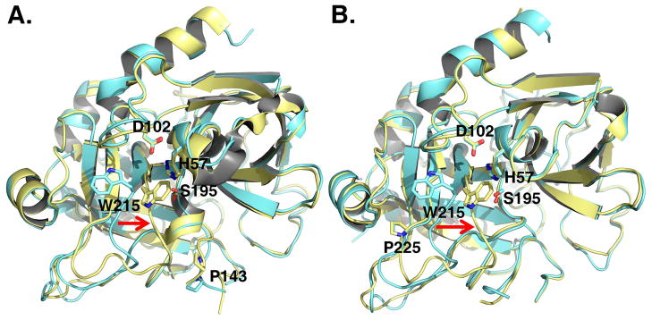
Figure 1. X-ray crystal structures of the thrombin mutants N143P and Y225P in the E* and E forms.
Ribbon representation of the structure of the thrombin mutants N143P (A) and Y225P (B) in the E (cyan) and E* (gold) forms. In the E* form the side chain of W215 and the entire 215–217 segment collapse into the active site (red arrow). In the E form, W215 moves back 10.9 Å and the 215–217 segment moves 6.6 Å to make the active site accessible to substrate. The rmsd between the two forms is 0.328 Å (N143P) or 0.345 (Y225P). Relevant residues are labeled and rendered as sticks.

