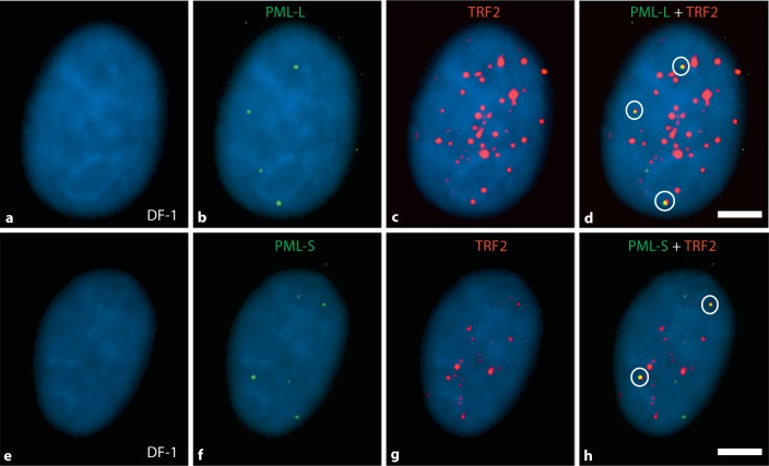
Fig. 4.
PML and TRF2 proteins in chicken nuclei: evidence for APB foci. Immunofluorescence detection of PML-L, PML-S, and TRF2 proteins was conducted using chicken-specific antibodies to detect colocalization of PML/TRF2 as an assay for the presence of ALT. Representative results are shown here for the DF-1 cell line. Images a–d show 1 cell from an experiment employing PML-L (green) and TRF2 (red); images e–h show a cell from an experiment employing PML-S (green) and TRF2 (red). Images d and h show merged images of all colors illustrating colocalization of PML-L or PML-S with TRF2, i.e. APB foci (yellow signals, 3 in image d and 2 in image h). Signal counts were conducted for PML-L, PML-S, and TRF2, and the data are shown in table 3. Colocalization of the PML and TRF2 (APB foci) was determined for each of the cell lines, and the results are shown in figure 5. Scale bar = 5 μm.

