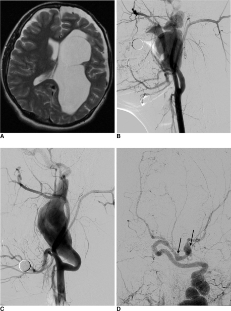
Fig. 1.
A. T2-weighted image of the patient's brain MRI shows porencephalic change of left cerebral hemisphere shows some cavitation suspiciously communicating with left lateral ventricle.
B. Angiography of the right ICA reveals large fusiform aneurysm arising from proximal ICA. The continuous draining artery shows marked tortuous dilation. The aneurysmal filling is not complete enough for the precise deliniation of whole contour of aneurysm.
C. Angiography of the left ICA reveals another giant fusiform aneurysm arising from the similar location to right side.
D. Intracranial view of left ICA angiography shows two more intracranial fusiform aneurysms (arrows) arising from proximal M1 segment and MCA bifurcation.

