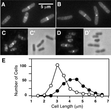
FIG. 3.
Localization of FtsE and FtsX to the division site. (A to D) Cells in exponential growth in LB with NaCl were fixed and examined by fluorescence microscopy directly (A and B), by indirect immunofluorescence microscopy (C and D), or by phase-contrast microscopy (C′ and D′). Strains shown are EC1063 (P204-ftsX-gfp) (A); EC1065 (P206-gfp-ftsX) (B); DHB4/pDSW609 (Plac-ftsE-3xHA) (C and D). (E) Relationship between cell length (age) and septal localization of FtsX. 509 cells of EC1063 were measured and scored for the presence (closed symbols) or absence (open symbols) of a fluorescent band at the midcell.

