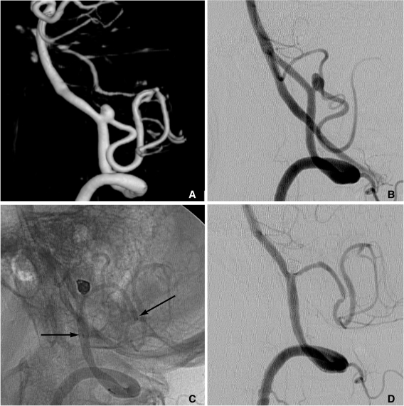
Fig. 18.
A, B. Images of 3D reconstruction and working projection show a wide necked aneurysm at the left VA-PICA origin.
C. Stent-assisted coiling is performed. Arrows indicate proximal and distal markers of the Enterprise stent.
D. The final control angiogram reveals complete occlusion of the aneurysm sac and widening of VA-PICA angle.

