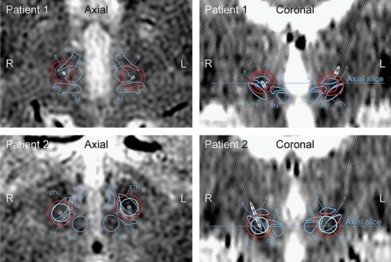
Fig. 3.
Axial and coronal images of group A (patients 1 and 2). These patients suffered from substantial stimulation-induced impairment of speech intelligibility during high-amplitude stimulation (i.e. 4 V). These patients had at least 1 of the active electrode contacts located in the posterior part of the STN. The boundaries of the red nucleus (RN), the STN, and the fct are marked in the axial and coronal images when present. The contours of the electric field isolevels are traced and color-coded where red indicates substantially decreased speech intelligibility and white indicates no effect on speech intelligibility.

