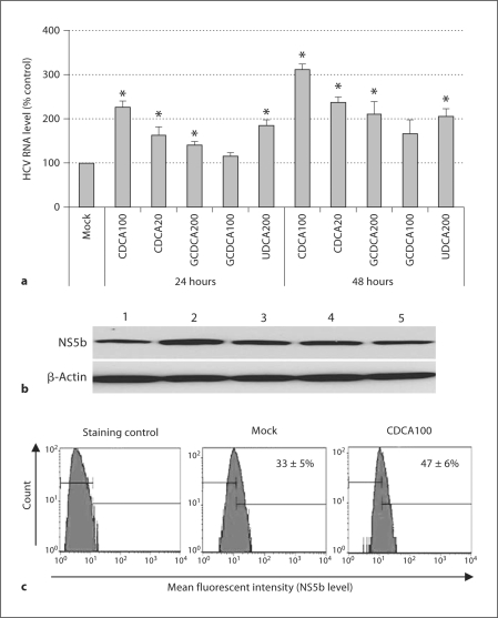
Fig. 1.
Enhancement of HCV replication after bile acid treatment in 1A7 cells. Semiconfluent cells were treated with mock medium, CDCA, GCDCA, or UDCA for 24 or 48 h. HCV RNA (a) or NS5b (b) was measured by real-time qRT-PCR or Western blot analysis, respectively. a qRT-PCR levels after treatment with mock medium, CDCA 100 μM (CDCA100), CDCA 20 μM (CDCA20), GCDCA 200 μM (GCDCA 200), GCDCA 100 μM (GCDCA100) and UDCA 200 μM (UDCA200) for 24 or 48 h. Asterisk (∗) indicates that the RNA levels by the treatment were significantly increased compared to those by control (mock medium) treatment (p < 0.05). b Western blot analysis detecting NS5b after the treatment for 24 h. Upper panel, lane 1: mock medium; lane 2: CDCA (100 μM); lane 3: CDCA (20 μM); lane 4: GCDCA (200 μM); lane 5: GCDCA (100 μM). Lower panel, as a loading control, Western blot analysis of β-actin was performed with the same samples. c Flow cytometry analysis of NS5b levels in 1A7 cells with the treatment of mock medium or CDCA (100 μM) for 24 h. Staining control was prepared using the same procedure without the incubation with the NS5b antibody.

