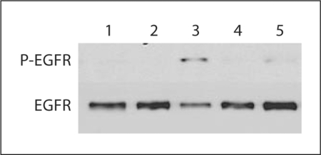
Fig. 3.
The activation of EFGR by the treatment with bile acids in GS4.1 cells. Confluent GS4.1 cells were treated with mock medium, CDCA, GCDCA, or UDCA for 30 min, and then cell lysates were prepared and Western blot analysis detecting phosphor-EGFR or EGFR. Lane 1: parental Huh-7 cells with mock treatment; lane 2: mock treatment; lane 3: CDCA 100 μM; lane 4: GCDCA 200 μM; lane 5: UDCA 200 μM.

