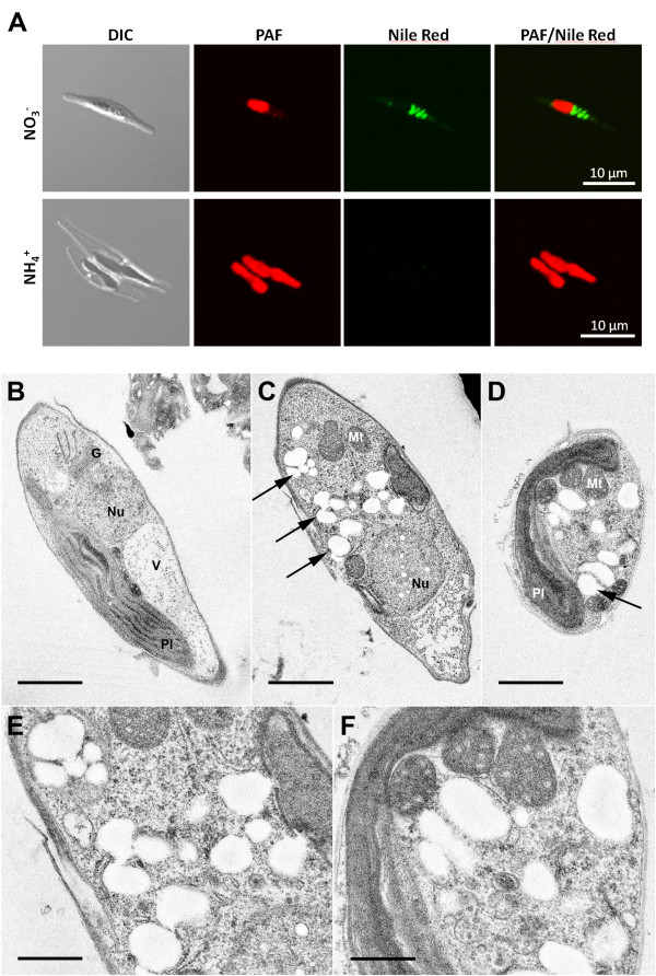
Figure 2.
Fluorescence and electron microscopic analyses on PHB accumulation in P. tricornutum. Cytosolic expression of enzymes PhaA, PhaB and PhaC of R. eutropha H16 induces the formation of granule-like structures that are stained by the lipophilic dye Nile red as visualized by fluorescence microscopy (A: NO3-). Under non-induced conditions no such granules were observed (A: NH4+). Electron microscopic analyses confirm cytosolic accumulation of electron-translucent granules (exemplarily marked by arrows) in cell lines expressing bacterial enzymes of the PHB pathway (C-F). PHB granules are about 0.1-0.3 μm in size and were not observed under non-induced conditions (B). Scale bar represents 1 μm (B-D) and 500 nm (E/F). DIC - differential interference contrast, G - golgi apparatus, Mt -mitochondrium, Nu - nucleus, PAF - plastid autofluorescence, Pl - plastid, V - vacuole

