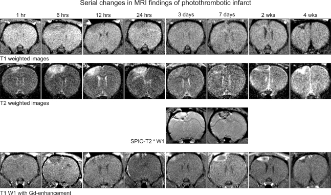
FIG. 2.
Early ischemic lesion (6-24 hours) revealed gradually decreased signal intensity of T1 weighted images, while T2 weighted images showed progressive increase of signal intensities. SPIO-T2 images defined macrophages infiltrations around necrosis as dark area between 3-7 days. Gadolinium-enhanced T1 weighted images show the enhancement in the area of fibrosis at 7 days- 4 weeks.

