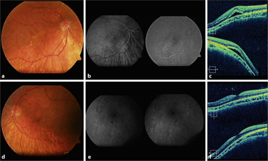
Fig. 1.
a–d Color retinography of both eyes in which posterior staphyloma and tilted disc are evident. b–e Fluorescein angiography showing retinal epithelial disturbance within the inferior and temporal macular area of the right eye; the left eye shows no abnormality. c Optical coherence tomography evidences dome-shaped macula and serous macular detachment in the right eye. f Optical coherence tomography evidences the presence of dome-shaped macula without subretinal fluid in the left eye.

