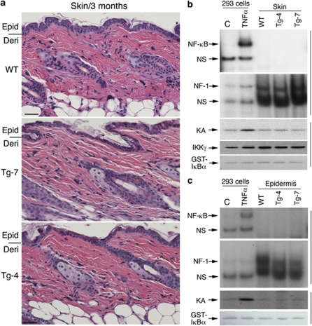
Figure 2.
No elevated IKK/NF-κB activation in the skin of mice overexpressing different levels of transgenic IKKα. (a) Histology of the skin from 3-month-old mice, stained with hematoxylin and eosin. Lines on the left of the panel indicate the division between the epidermis and dermis. Epi, epidermis; Der, dermis. Scale bars=150 μm. (b) NF-κB and IKK kinase activity in the skin, and (c) epidermis was detected using gel shift assay and immunoprecipitation kinase assays. HEK 293 cells treated with TNFα (10 ng/ml) for 20 min were used as the positive control. Glutathione S-transferase-IκBα, stained with Ponceau S solution, was used as a substrate of IKK. Antibody against IKKγ was used to precipitate the IKK complex. NF-1, sample loading control; NS, non-specific band

