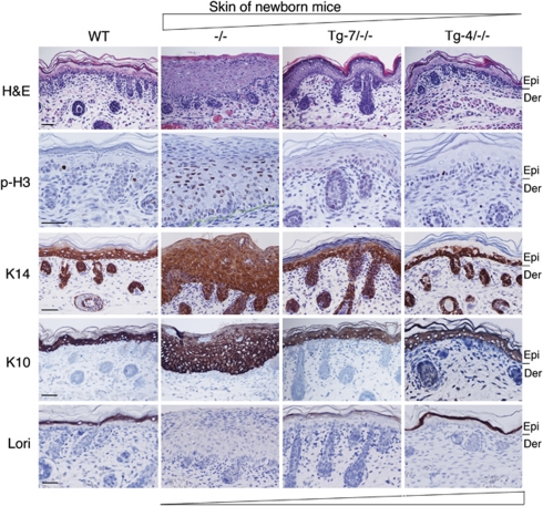
Figure 4.
Correlation of transgenic IKKα levels with epidermal thickness and differentiation. Paraffin-embedded skin sections of newborn mice were stained with hematoxylin and eosin, or immunohistochemically stained with phosphorylated histone H3 (p-H3), K14, K10, and loricrin (Lori). The angle sign at the top of the panel indicates the reduced thickness of the epidermis and mitotic activity. The angle sign at the bottom of the panel indicates the increased terminal differentiation and IKKα doses. The lines on the right of the panel and the green lines in the p-H3-stained skin of Ikkα−/− mice (−/−) indicated the division between the epidermis and dermis. Dark brown color indicates positive staining; blue color indicates hematoxylin counterstaining. Epi, epidermis; Der, dermis; −/−, Ikkα−/−. Scale bars=150 μm

