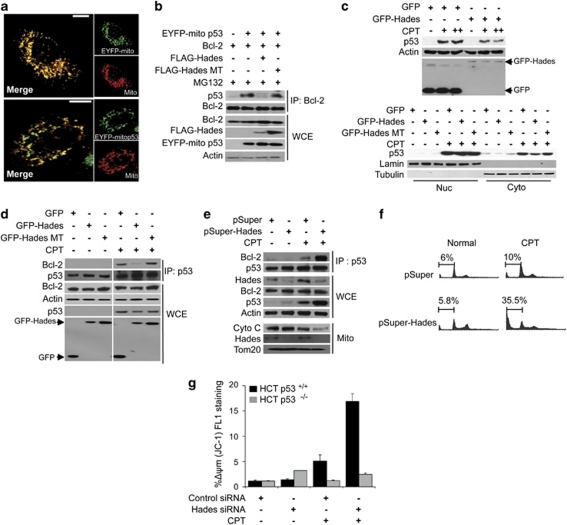
Figure 5.
Hades regulates the exonuclear function of p53. (a) Confocal microscopic images of pEYFP-mito p53 localization in U2OS mitochondria at 24 h after transfection with mitotracker and pEYFP-mito p53. (b) Hades inhibits interaction between p53 and Bcl-2. HEK293 cells were cotransfected with expression plasmids for EYFP-mito-p53, Bcl-2, FLAG-Hades, and FLAG-Hades MT as indicated. The cell lysate was immunoprecipitated with anti-Bcl-2 antibody, then immunoblotted using anti-p53 antibody. (c) Hades attenuates camptothecin (CPT)-induced increases in p53. MCF7 cells were transfected with expression plasmid for GFP-Hades or GFP-Hades RING MT. At 24 h after transfection, cells were treated with CPT (1 or 5 μM (upper), 5 μM (lower)) for 6 h. The p53 protein level was analyzed by immunoblotting in whole-cell extracts (upper) and in nuclear and cytoplasmic fractions (lower). Anti-Lamin and anti-Tubulin antibodies were used as fractionation and loading controls, respectively. (d) Hades inhibits interaction between p53 and Bcl-2 in CPT-treated cells. MCF7 cells were transfected with expression plasmids for GFP-Hades or GFP-Hades MT for 24 h then further incubated with 1 μM CPT for 24 h. Anti-p53 immunoprecipitates were immunoblotted using anti-Bcl-2 and anti-p53 antibodies. (e) Stable Hades knockdown stimulates the interaction between Bcl-2 and p53 following 1 μM CPT treatment for 24 h in MCF7 cells. Immunoprecipitate complexes using anti-p53 antibody were subjected to immunoblotting as in (d) (upper). Whole-cell extracts were probed for p53, Hades, and Bcl-2 (center), whereas mitochondrial fractions were probed for cytochrome c (lower). (f) Hades ablation increases apoptosis among CPT-exposed cells. Stable Hades-knockdown MCF7 cells were exposed to 1 μM CPT for 24 h, stained with propidium iodide, and analyzed by FACS to measure the sub-G0 fraction. (g) siRNA-induced knockdown of Hades promotes CPT-induced mitochondrial damage in a p53-dependent manner. HCT116 p53+/+ and HCT116 p53−/− cells were transfected with 40 nM Hades siRNA or control siRNA, incubated for 24 h, exposed to 1 μM CPT for 24 h, stained with JC-1 for 30 min, and analyzed by flow cytometry. Error bars represent the means (S.E.M.) of at least three independent experiments

