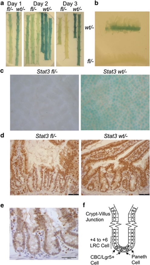
Figure 1.
Use of a Flox-STOP LacZ transgene as a surrogate marker of Stat3fl recombination to visualise the transient appearance and disappearance of STAT3-null crypts. (a) LacZ enzyme activity in Stat3fl/− and Stat3wt/− small intestine at 1, 2 and 3 days after β-naphthoflavone injection/induction of Cre expression. (b) Cross-section of small intestine showing LacZ enzyme activity in Stat3fl/− and Stat3wt/− mice at 3 days after β-naphthoflavone injection/induction of Cre expression. (c) Crypt bases in Stat3fl/− and Stat3wt/− small intestine at 3 days following β-naphthoflavone injection/induction of Cre expression viewed at high power ( × 90 magnification) after removal of the smooth muscle layer. LacZ enzyme activity panels are representative of three Stat3fl/− versus three Stat3wt/− mice per time point. (d) Anti-STAT3 immunoreactivity (with the NEB/CST #9132 primary antibody recognising an epitope close to the STAT3 tyrosine 705 residue) in Stat3fl/− versus Stat3wt/− small intestine at 1.7 days after induction of Cre expression. Immunohistochemistry panels are representative of three Stat3fl/− versus three Stat3wt/− mice, the black bars represent 50 μm. (e) Anti-STAT3 phosphotyrosine 705 immunoreactivity (using the NEB/CST #9131 primary antibody) in un-induced Stat3wt/− control small intestine. The panel is representative of three Stat3wt/− mice, the black bar represents 50 μm. (f) Schematic of the small-intestine crypt base showing the thin CBC/Lgr5+ slowly proliferating stem cells interleaved by Paneth cells with the over-lying +4 to +6 region containing long-term, label-retaining, quiescent stem cells

