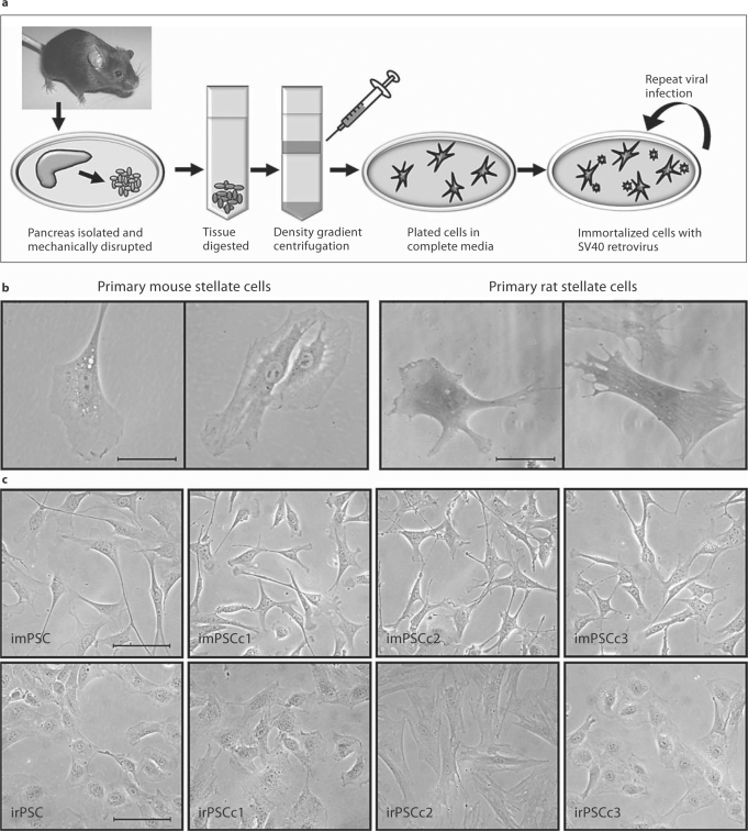
Fig. 1.
Methodology and resultant PSC isolation and immortalization. a As shown, PSC were isolated from rat and mouse. Briefly, the pancreas was isolated, mechanically disrupted, enzymatically digested, and stellate cells isolated by Accudenz gradient centrifugation. b Microscopy images of primary rat and mouse stellate cells, with some cells containing vitamin-A-containing vacuoles. Scale bar represents 50 μm. c To establish immortalized cell lines and clones, primary stellate cells were infected with ecotropic virus containing the SV40 large T antigen. From a heterogeneous population of cells, three clonally expanded cell lines were established for each the mouse and rat pancreas. Phase images of heterogeneous and clonally expanded imPSC and irPSC clones 1, 2, and 3. Scale bar represents 100 μm.

