Fig. 2.
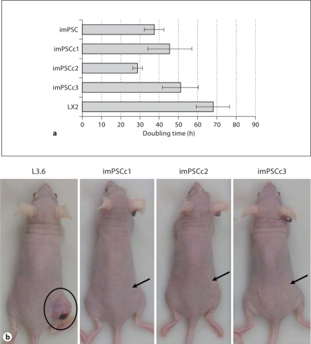
PSC lines have distinct in vitro and in vivo growth characteristics. a PSC rapidly divided and doubled every 24–48 h. Hepatic (LX2) and pancreatic (imPSC) stellate cells were plated and incubated for up to 72 h. At 24-hour intervals, cells were trypsinized, trypan blue stained and manually counted with a hemacytometer. Exponential best-fit curves from three independent experiments were averaged and doubling time calculated ± SEM. b Immortalized PSC were unable to establish subcutaneous tumors in nude mice. Athymic nu/nu mice were injected with control L3.6 and immortalized PSC lines and observed after 3 weeks. Tumor formation was evident in L3.6 mice (837.8 mm3) and essentially absent in all imPSC clones (imPSCc1, none; imPSCc2, 9.4 mm3; imPSCc3, none). Arrows indicate injection site on mouse flanks.

