Fig. 3.
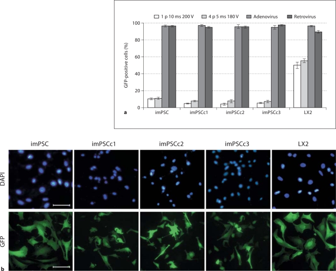
Immortalized mPSC efficiently expressed exogenous GFP after adenoviral and retroviral transductions. Stellate cells were subjected to a variety of transfection and transduction protocols to determine the ability of these cells to express exogenous GFP. Cells expressed GFP 48 h after electroporation, at two different voltages and pulse rates, or incubation with adenovirus or retrovirus. a Cells were counted (100–500 per replicate) and percentage of total population expressing GFP calculated, n ≥ 3 independent replicates. b Representative images of an adenoviral infection of the stellate cells and clones. Scale bar represents 100 μm.

