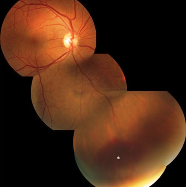
Fig. 2.

Fundus photograph taken after the resolution of the exudative retinal detachment induced by the TASER. The resolving subretinal hemorrhage, as well as the adjacent smaller retinal hemorrhages, and pigmented scars are seen (asterisk). The montage shows the size and location of the detachment in relation to the posterior pole
