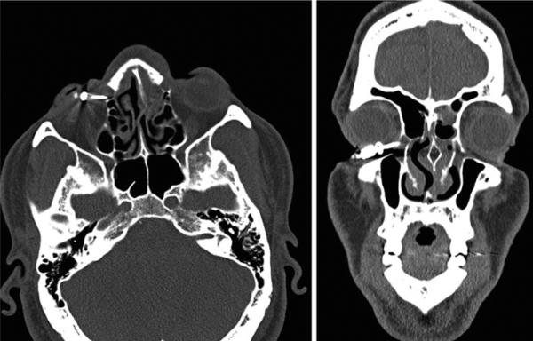
Fig. 3.
(Left) Axial view of the orbital CT scan showing the tip of the TASER dart in the right lacrimal fossa. (Right) Coronal section of the orbital CT scan. No evidence of globe or muscular lesion. The hemorrhagic fluid in left ethmoidal cells is the result of a broken nose, from a fall sustained after being “TASERed”

