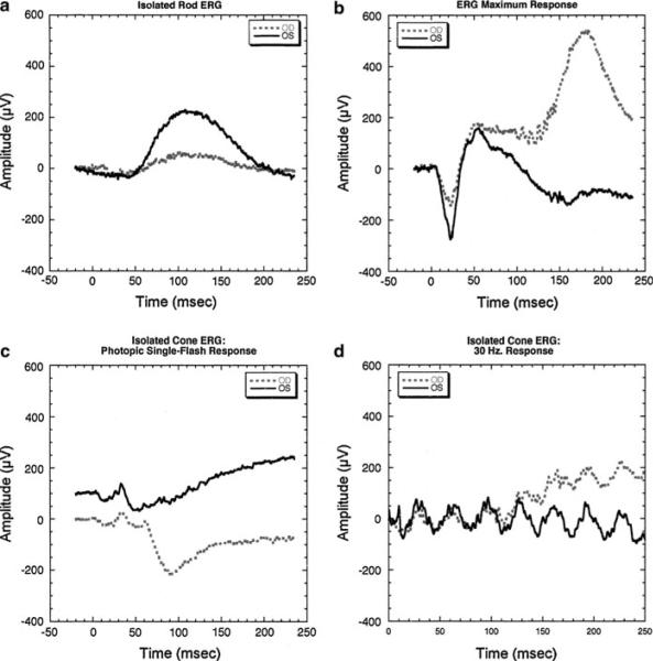
Fig. 5.
Ganzfeld electroretinogram performed 3 days after the TASER injury. a Isolated rod responses of the affected eye (OD; gray dashed line) compared to the concomitant response from the normal eye (OS; dark solid line). There is a 70% reduction in the b-wave amplitude of the affected relative to the fellow eye. b The ERG maximum response shows about a 63% reduction in the rod photoreceptor response, consistent with the b-wave reduction in a, and suggesting that the b-wave reduction results from reduced photoreceptor responses. The reduction in ERG amplitude in a and b is greater than what would be expected from the extent and location of the observed exudative retinal detachment. c Isolated cone responses to a single flash of light. Responses are vertically displaced for comparison. The response from the affected eye is reduced 10% when compared to the normal eye and is minimally delayed by 1–2 ms. d Isolated cone responses to a 30-Hz flickering light reflect the minimal amplitude and timing changes observed in c

