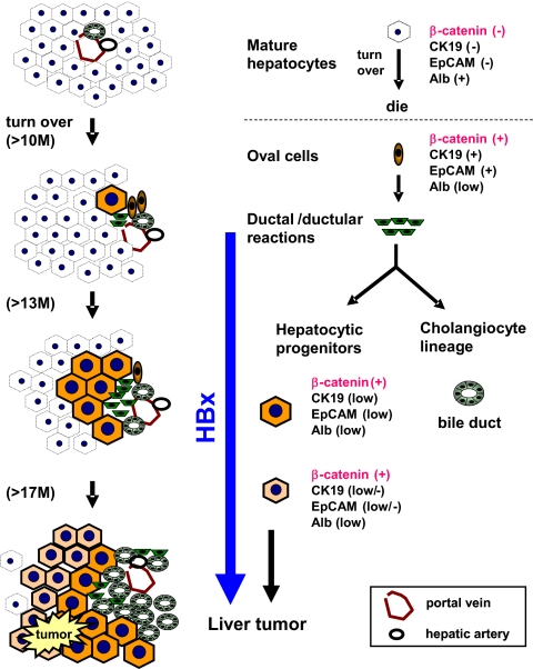
Fig. 5.
Schematic illustration of the replacement and carcinogenic events in the liver of β-catenin–KO mice, showing changes in cell populations at different ages. The liver consisted of approximately 99% β-catenin(−) mature hepatocytes from day 15 after birth for the normal hepatocyte lifespan (>10–12 mo), after which the hepatocytes underwent regeneration. Because hepatocytes lacking β-catenin lose the ability to regenerate, hepatic oval cells proliferate and differentiate into hepatocytes and cholangiocytes. These two cell lineages gradually replaced the entire liver mass. During active proliferation, most hepatic progenitor cells underwent maturation arrest and became dedifferentiated via an unknown mechanism. These progenitor-derived immature hepatocytes showed a high potential to develop into liver tumors, including adenoma and HCC, and this process was accelerated by HBx.

