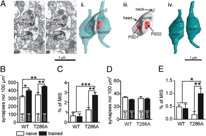
Fig. 2.
Training-induced generation of MIS in T286A mutants but not in WT mice. (Ai) Two serial electron micrographs with a single spine innervated by two axonal boutons (axon1 and axon2), each with single postsynaptic density (PSD1 and PSD2). (Aii) 3D reconstructions of this MIS. The red color indicates the PSDs contacting the spine. (Aiii) The spine without the axonal boutons. (Aiv) Two axonal boutons. Synapse density and MIS were analyzed 2 h after conditioning in stratum radiatum of hippocampal area CA1 by using 3D electron microscopy (B and C) and 24 h after training by using 2D electron microscopy (D and E).

