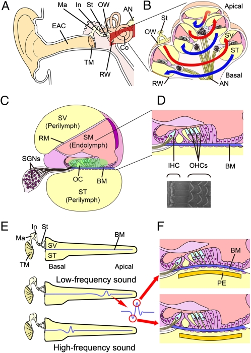
Fig. 1.
Anatomy of the mammalian cochlea. (A) Schematic drawing of the human auditory system. AN, auditory nerve; Co, cochlea; EAC, external auditory canal; In, incus; Ma, malleus; OW, oval window; RW, round window; St, stapes; TM, tympanic membrane. (B) Schematic drawing of a cochlear coil. Sound vibrations transmitted to the cochlea fluid in the scala vestibule (SV) through the OW travel up from the basal turn to the apical turn (red lines) and then back to the basal turn (blue lines) in the scala tympani (ST). (C) Schematic drawing of a cochlear duct. The ST and SV are filled with the perilymph. The scala media (SM), which is separated from the ST by Reissner's membrane (RM) and separated from the ST by the basilar membrane (BM), is filled with the endolymph. The organ of Corti (OC) is located on the BM. Spiral ganglion neurons (SGNs) are located in the modiolus of the cochlea. (D) Schematic drawing and scanning EM of the organ of Corti. One row of inner hair cells (IHCs) and three rows of outer hair cells (OHCs) are arranged along the entire length of the cochlear coil. (E) Schematic drawing of traveling wave theory. Low-frequency sounds vibrate the BM in the apical portion of a cochlea, whereas high-frequency sounds provoke vibration in the basal portion of a cochlea. (F) Schematic drawing showing the vibration of the BM and a piezoelectric membrane (PE) implanted in the ST of a cochlea in response to sound stimuli.

