Fig. 1.
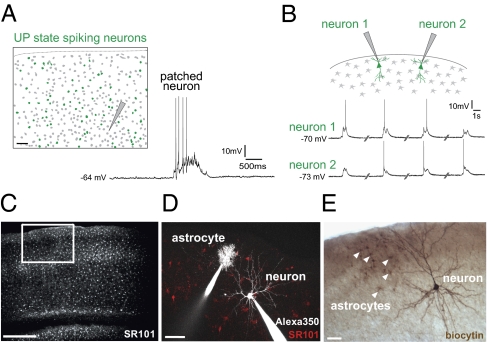
UP states are synchronous, network-wide events. (A) Cortical slice labeled with Fura-2. Cells in green displayed significant calcium increases, corresponding to action potentials, during the UP state recorded in a layer 5 neuron (patched). Gray cells did not change in Fura-2 fluorescence and presumably did not fire action potentials. (Right) Recorded UP state. (B) Neurons 500-μm apart depolarize synchronously during UP state. Traces are not continuous in time. We never observed an UP state in one neuron but not in the other. Note that each neuron depolarizes during the UP state but does not always spike. (C) Two-photon image of astrocytes across all six cortical layers in area S1, labeled with SR101. The white box represents the extent of cortical layers for subsequent imaging. (D) Whole-cell patch-clamped astrocyte (layer 1) and pyramidal neuron (layer 2/3) with Alexa 350 (white) in pipettes. Astrocytes labeled with SR101 (red). (E) Biocytin-labeled neuron and astrocytes, five of which are marked by arrowheads. These are not the same two cells as in D. (Scale bars, 50 μm.)

