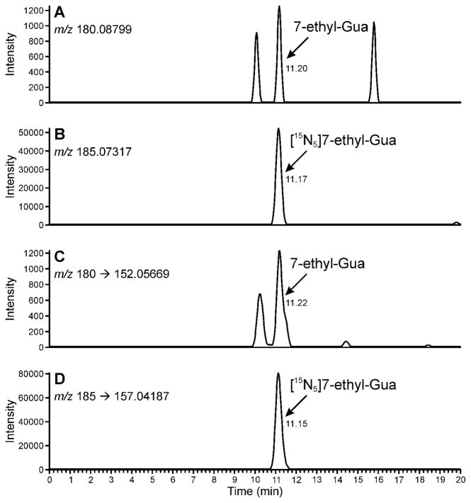
Figure 2.
Chromatograms obtained upon LC-NSI-HRMS/MS-SRM analysis of human leukocyte DNA (129 μg, 12.9 μg on column) containing 59.4 fmol/μmol Gua. The relatively higher amount of analyte in this sample allowed confirmation of its identity by additional monitoring of the accurate mass of the molecular ion of 7-ethyl-Gua and the internal standard. Panel (A) shows the result from monitoring of the accurate mass of 7-ethyl-Gua (m/z 180.08799). Panel (B) shows the result from the monitoring of the accurate mass of [15N5]7-ethyl-Gua (m/z 185.07317). Panel (C) shows the results from the transition at m/z 180 [M + H]+ → m/z 152.05669 [Gua + H]+ for 7-ethyl-Gua and panel (D) shows the corresponding transition m/z 185 [M + H]+ → m/z 157.04187 [Gua + H]+ for the internal standard. Results are shown with a 5 ppm mass tolerance.

