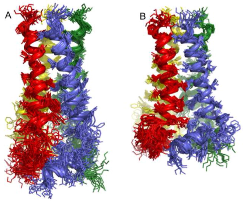
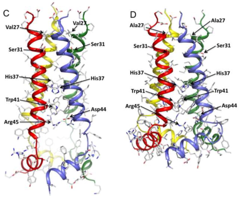
Figure 2. Structures of WT and the V27A mutant.


a. Superposition of 15 low energy structures of WT18-60 (2RLF)[16] and b. V27A18-60 (2KWX). The V27A structures were calculated using restraints summarized in Table 1. c. Ribbon representation of the WT structure (2RLF) and d. the V27A structure (2KWX). Compared to WT, the structure ensemble of V27A shows better-defined arrangement of the AP helices relative to the pore domain due to more long-range NOEs.
