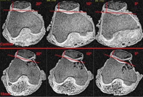
Figure 5.
Images were collected using dynamic MRI during the final 20 degrees of knee flexion. When comparing me control and 15mm-transfer knees, it is clear that me amount of patella lateral to me perpendicular drawn from me apex of me lateral femoral condyle is reduced. Additionally, in both the control and 15mm knee, the amount of patella lateral to the perpendicular increases as the knee nears extension (0 degrees flexion).

