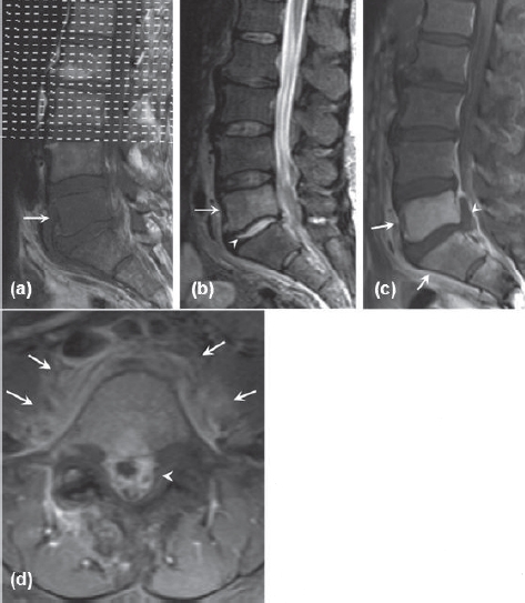
Figure 8.

Initial MRI for patient two. (a) Sagittal T1-weighted image showing hypointense lesion in the L5 vertebral body. (b) Sagittal T2-weighted image with hyperintense lesion in the L5 vertebral body and hyperin-tense L5-S1 disk. (c) Sagittal Tl-weighted post-gadolinium image with fat suppression showing L5 and S1 vertebral body enhancement (arrows) and epidural abscess (arrowhead). (d) Axial T1weighted post-gadolinium image showing epidural (arrowhead) and paravertebral (arrow) enhancement
