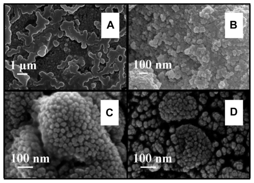
Figure 4.
FESEM images of nano-HAP powder samples at various thermal treatment temperatures (scale bars shown in each image). (i) No ultrasound in preparation (A) 300°C and (B) 400°C (ii) Ultrasound used in preparation (C) 300°C and (D) 400°C.
Abbreviations: FESEM, field emission scanning electron microscopy; HAP, hydroxyapatite.

