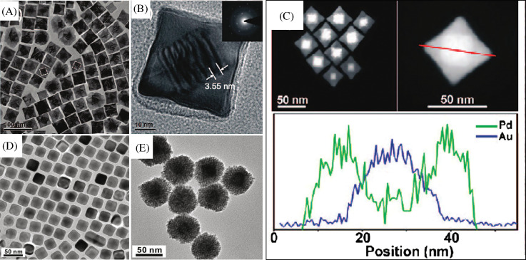
Fig. 4.
(A) TEM image of Au@Pd nanocubes. (B) TEM image of a single Au@Pd nanocube at high magnification. The inset is the SAED pattern taken from individual nanocube. (C) STEM images of the octahedral Au seed within a cubic Pd shell and cross-sectional compositional line profiles of a Au@Pd nanocube along the diagonal (indicated by a red line). D and E are TEM images of Au@Ag nanocubes and Au@Pt nanoparticles, respectively. Reproduced with permission from Reference (166). American Chemical Society, Copyright (2008).

