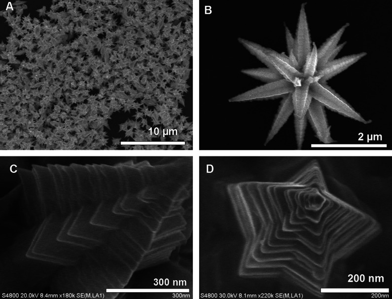
Fig. 8.
Large area (A) and corresponding single particle. (B) Field-emission scanning electron microscopy (FESEM) images of gold MFs. (C) An enlarged FESEM image of a single stem of the MF showing ridges along the edges. (D) Top view of a single stem of the MF showing the pentagonal structure. Reproduced with permission from Reference (169). Springer, Copyright (2009).

