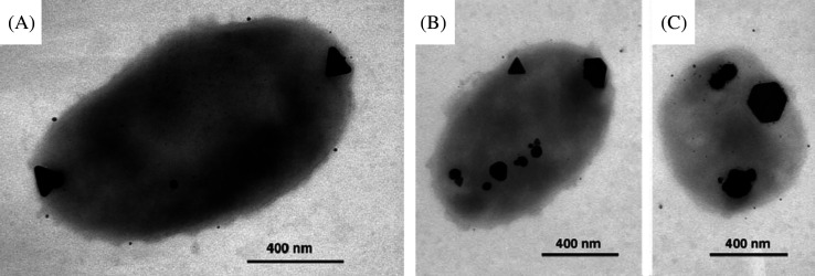
Fig. 15.
(A) TEM image of large, triangular, Ag-containing particles at both poles produced by P. stutzeri AG259. An accumulation of smaller Ag-containing particles can be found all over the cell. (B and C) Triangular, hexagonal, and spheroidal Ag-containing nanoparticles accumulated at different cellular binding sites. Reproduced with permission from Reference (186). Proceedings of National Academy of Science, Copyright (1999).

