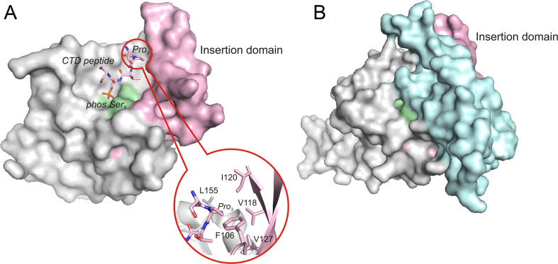
Fig 5.
Surface representation of Scp1 and Fcp1 FCPH domains. (A) Scp1 in complex with a short CTD peptide (2ght). The zoom-in picture shows the Pro3 binds to the hydrophobic pocket with the aromatic residues shown in stick. (B) Fcp1 FCPH domain (3ef0). In both structures, the active site signature motif is colored with pale green, and the insertion domain is colored with light pink. Notably, the additional helical domain (light cyan) covers the ‘insertion domain’ in Fcp1 and makes it much less accessible for substrates.

