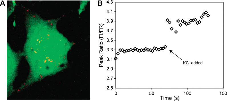
Fig. 10.
(A) Compressed z stack of confocal image of a single C6 glioma cell injected with magnesium-selective PEBBLE probes (Type 2 probe). The probes, depicted in red, reside in the cytosol and do not enter the nucleus at the center of the cell. (B) Spectra of the intercellular probes acquired on a fluorescence microscope. An aliquot of KCl was added at t=69 s. Ion channels open, resulting in an increase in intracellular free magnesium indicated by the increase in peak ratio of the probes. Adapted from Reference (116). Reprinted with permission from ACS Publishing Group, Copyright (2003).

