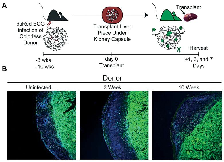
Fig. 2.
Transplantation of uninfected, 3- and 10-week BCG infected colorless liver tissue under the kidney capsule of GFP recipient mice. A, Experimental Scheme. B, Fluorescent microscopy of kidney sections 3 days post transplant of uninfected, 3- and 10-week BCG infected colorless liver tissue. Images taken at ×10 magnification. GFP (green) and DAPI nuclear stain (blue)

