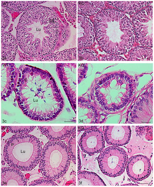
Figure 3.
Hematoxylin and eosin stained adult testis (a,b), efferent ducts (c,d) and proximal initial segment (e, f) of 10–12 month old wildtype (WT, Cst8+/+) (a,c,e) and knockout (KO, Cst8−/−)(b,d,f) mice. Tubules in Cst8−/− mice were consistently smaller compared to WT. SE, seminiferous epithelium; Lu, lumen. Bars: a,b,e,f = 50 μm; c,d = 20 μm.

