Figure 5.
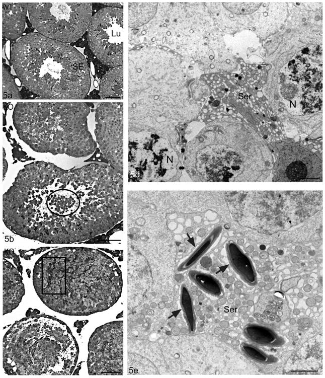
Light micrographs of the seminiferous epithelium of WT (Cst8+/+) (a) and KO(Cst8−/−) (b,c) 10–12 month old mice. In comparison to the intact seminiferous epithelium and unobstructed tubular lumen of WT mice (a), Cst8−/− tubules show shedding of immature germ cells into the lumen (b, circle) and disoriented late spermatids (c, box). Cst8−/− mice also display vacuolization in the epithelium, closure of the tubule lumen (b) and detachment of germ cells at the junction of the basal/adluminal compartment (c, asterisks). Electron micrographs (d,e) of the seminiferous epithelium of Cst8−/− 10–12 month old mice. In (d), several germ cells adjacent to a Sertoli cell (Ser) show nuclei (N) with abnormal chromatin patterns and pale stained areas including membranous whorls, all suggestive of degeneration. In (e), several late elongating spermatids are closely enveloped by a Sertoli cell (Ser) and although they have an acrosome, they are not oriented properly and show no association with ectoplasmic specializations. An empty looking space is present surrounding these spermatids (arrows). Seminiferous epithelium, SE; Lumen, Lu. Bars: a,b,c = 50 μm; d,e = 2 μm.

