Figure 7.
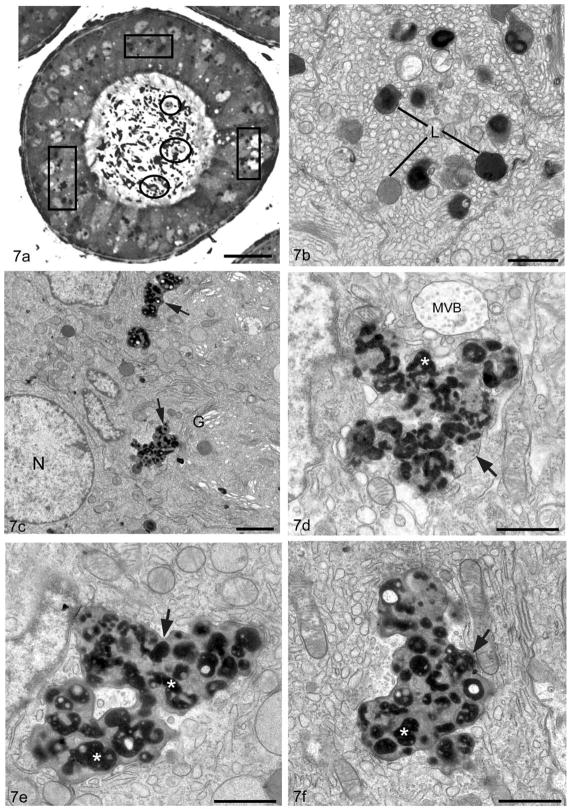
Light micrograph (a) of a tubule of the proximal initial segment of a 10–12 month old Cst8−/− mouse displays large irregularly shaped supranuclear lysosomes (boxes) in principal cells and immature germ cells in the epididymal lumen (circles). Electron micrographs of the supranuclear region of principal cells of the initial segment of a WT (Cst8+/+)(b) and KO (Cst8−/−) (c–f) 10–12 month old mice. In WT (b), lysosomes (L) are small in size, more or less spherical and reveal dense granular whorls embedded in a moderately dense material or a homogeneous dense or moderately dense matrix. In KO (c–f), lysosomes (arrows) are extremely enlarged and pleomorphic in appearance. Surrounded by a unit membrane they contain numerous dense bodies (asterisk) with irregularly shapes and sizes in which paler areas are present; some dense bodies are at times spherical, bead-like or tubular in nature. Multivesicular body, MVB; nucleus, N; Golgi apparatus, G. Bars: a = 25 μm; b,d,e,f = 1 μm; c = 2 μm.

