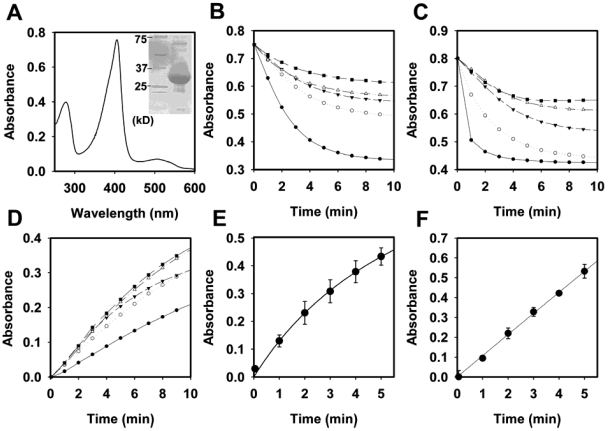
FIG 1 .
Biochemical and enzymatic characterization of YfeX. (A) UV/visible scan of heme-loaded purified YfeX, as described in the text. The heme Soret peak has a maximum at 415 nm. The inset shows a sodium dodecyl sulfate-polyacrylamide gel that is overloaded with protein to demonstrate the purity of the YfeX protein employed in these studies. (B) Alizarin red dye decolorization by YfeX. Details are in Materials and Methods. (C) Cibacron blue dye decolorization by YfeX. (D) Pyrogallol oxidation by YfeX. Symbols (B, C, and D): solid circles, pH 5.5; open circles, pH 6.0; solid triangles, pH 6.5; open triangles, pH 7.0; and solid squares, pH 7.5. (E) Protoporphyrinogen IX oxidation to protoporphyrin IX by YfeX. (F) Coproporphyrinogen III oxidation to coproporphyrin III by YfeX.

