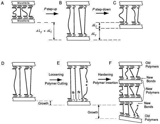
Figure 9.
Diagrammatic representation of elastic changes and growth in a molecular unit of the primary wall. A, Cellulose microfibrils connected through hydrogen bonds (>>>>) to tethering matrix polysaccharides, two of which are load-bearing. B, More tethers become load-bearing (ΔLr) and some tethers are displaced (ΔLi) when P increases. C, Some tethers are released from load-bearing but displacements are not fully reversed when P decreases. D, Wall at high P (as in B) is loosened in E by breaking covalent bonds (★) and hydrogen bonds (⋆) probably through enzymatic action. F, Inserting new wall polymers and forming new hydrogen bonds hardens the wall in E. Not shown are wall proteins or layering of various polysaccharides in the wall.

