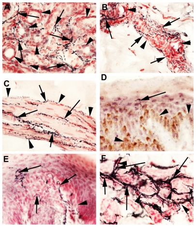
Figure 5.
SV2A in human skin. SV2A-immunoreactive fibers (arrowed) in sweat glands (arrowheads) (A), around blood vessels (arrowheads) (B), arrector pili (C), and rare intraepithelial fiber, epithelial basal layer indicated by arrowhead (D) in human skin from a patient with small fiber neuropathy. Rat paw skin showing intraepithelial SV2A-immunoreactive fibers, epithelial basal layer indicated by arrowhead (E) and dense immunoreactive fibers around sweat glands (arrowheads) (F), magnification 40×.

