Fig. 1.
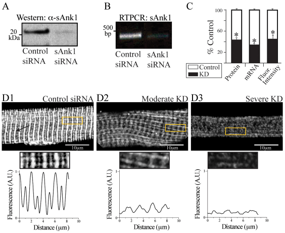
Effect of targeted siRNA on sAnk1 mRNA and protein levels. FDB fibers were transduced with adenovirus encoding siRNA selective for sAnk1 or a control sequence. Samples were maintained in culture for 48 hours and then examined by western blotting, RT-PCR and immunofluorescence. (A,B) sAnk1 protein (A) and mRNA (B) are reduced in cultures of fibers treated with sAnk1-siRNA. (C) Mean sAnk1 protein, cDNA and fluorescent intensity levels are significantly reduced (>50%) in cells treated with sAnk1-siRNA (KD), compared with controls (*P<0.01, Student's t-test). Results are means ± s.e.m. (D) In con-siRNA-treated myofibers (D1), sAnk1 concentrates around Z-disks, and to a lesser extent around M-bands, as indicated by the regular fluorescent peaks. Myofibers treated with sAnk1-siRNA (D2, D3), show a range of intensities and distribution, from little (similar to controls; not shown), to moderate (D2) and severe (D3); 5× regions of interest (ROIs) boxed in yellow. Severely affected fibers (D3) constitute ~15% of the total. Most fibers are only moderately affected (D2), and show reduced labeling for sAnk1, that is absent around the M-band, and present in some longitudinal structures. The arrow in D1 indicates Z-disks. See also supplementary material Fig. S1. The results show that sAnk1-siRNA reduces sAnk1 levels and alters its distribution in skeletal myofibers.

