Fig. 2.
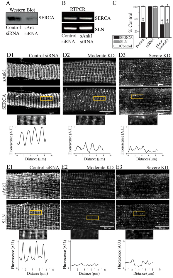
Reduced expression of sAnk1 leads to disruption of the network SR. FDB myofibers were treated as in Fig. 1 and analyzed for two components of the nSR, SERCA and SLN. (A) SERCA protein is significantly reduced in immunoblots of myofibers treated with sAnk1-siRNA, compared with con-siRNA. (B) Levels of mRNA encoding SERCA and SLN are not altered in myofibers in which sAnk1 expression is reduced. (C) Intensity of immunofluorescence labeling of SERCA and SLN are both significantly reduced (>50%) in cells treated with sAnk1-siRNA, compared with controls. Results are means ± s.e.m. (D,E) The organization of SERCA (D2,D3) and SLN (E2,E3), is disrupted or lost when sAnk1 expression is inhibited as compared with controls (D1 and E1, respectively). Fluorescent profiles of the ROI (yellow boxes) illustrate the normal, striated pattern of SERCA and SLN, which localize primarily at the Z-disks in control fibers (bottom left panels of D1 and E1, respectively), which is lost in fibers with very low levels of sAnk1 (bottom right panels of D3 and E3, respectively). The results show that sAnk1-siRNA reduces the amount of SERCA and SLN proteins, but not their mRNAs, and alters their distribution in skeletal myofibers.

