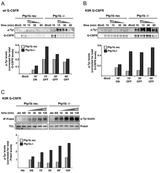
Fig. 6.
Increased tyrosine phosphorylation of G-CSFR and Prdx4 in Ptp1b-deficient cells. (A) Phosphotyrosine analysis of wild-type G-CSFR after biotinylated G-CSF (BioG) pull-down in Ptp1b−/− or Ptp1b reconstituted MEFs expressing wild-type G-CSFR (Ptp1b rec). Ten minutes after addition of biotinylated G-CSF, cells were washed and further cultured without growth factor. Histograms show quantifications of Tyr-P levels relative to total G-CSFR protein in biotinylated G-CSF pull-downs at the indicated times. Data shown are representative of three independent experiments. (B) Similar to A, with lysine-less G-CSFR mutant K5R that accumulates in early endosomes. (C) Phosphotyrosine analysis of Prdx4 immunoprecipitates in Ptp1b−/− or reconstituted control MEFs at various times after stimulation with G-CSF. Histograms show quantification of Tyr-P (Odyssey) levels relative to immunoprecipitated Prdx4. Data are representative of two independent experiments. TCL total cell lysates, NS not stimulated.

