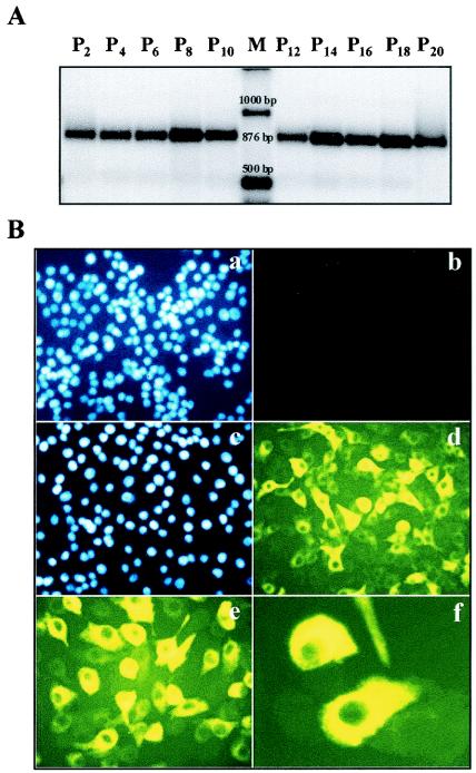
FIG. 1.
Characterization of an MDBK cell line persistently infected with ncp BVDV. (A) Viral RNA was purified from the culture supernatants harvested every 3 days when the cells were passaged and subsequently used to perform an RT-PCR analysis; 20% of the RT-PCR product was loaded and run on a 2% agarose gel stained with ethidium bromide. (B) Noninfected cells (panels a and b) and the MDBK cell line, at passage 5 after establishment, persistently infected with ncp-BVDV (panels c to f) were fixed and probed with monoclonal antibodies directed against BVDV E2 and NS2/3 proteins, followed by incubation with an anti-mouse-FITC-conjugated secondary antibody (d to f). Nuclear staining with DAPI was performed to quantify the cells (a and c).

